
Location
Montrose Regional Health
800 S. 3rd Street
Montrose, CO 81401
Appointments: 970-249-2211
Ambulatory Care Center
3330 S. Rio Grande Ave
Montrose, CO 81401
Appointments: 970-497-5976
Imaging Services in Montrose
MRH is committed to what our community needs most. Services like cancer care, women’s health and obstetrics require the best imaging service in the region. That’s why our Medical Imaging Department remains focused on continuing advancements in imaging – to bring you information that’s critical to your health.
Learn more about our radiology services by clicking on the toggles below.
Welcome to Breast Health, Imaging, & Procedures at Montrose Regional Health. Whether your needs are a routine mammogram or for diagnostic imaging and procedures to help gain insight, we’re here to help you.
The most effective way to detect early signs of breast cancer and address other noncancerous breast conditions is to undergo routine preventative care including mammograms.
Our team meets the highest standards for our patients while also providing them and their families support, comfort, and answers. You can ask questions and discuss concerns about your breast health comfortably and confidentially. To help you feel at home, we make every effort to accommodate requests for a specific technologist when scheduling your routine mammogram and other important breast exams and tests.
Mammograms
Breast cancer is the most common cancer in women. In today’s advanced medical environment, there is more hope than ever before for breast cancer patients. One of the keys is early detection; at MRH, we can perform these routine screening and diagnostic tests with efficient procedures and highly trained staff.
Today’s gold standard in breast cancer screening is 2D mammography. However, it doesn’t always deliver clear results. Traditional 2D mammograms suffer from decreasing sensitivity as the density of a woman’s breast tissue increases, or when breast tissue overlaps. This can result in unclear images that leave health providers unsure of what they see, and can lead to cancers being missed.
MRH offers True Breast Tomosynthesis, a state-of-the-art 3D mammogram technology that is proven to detect breast cancer better than traditional 2D mammography alone. We’re using it in our practice to give women a new level of confidence in their breast health.
Breast Lumps
In most cases, breast lumps are benign (noncancerous) and your provider will recommend monitoring them for changes. If a positive diagnosis is ever received, we are available for you throughout your treatment and recovery.
We can also provide our patients with information about the local Bosom Buddies organization to help connect you with even more support in your journey back to great health.
Breast Imaging Tests
• Breast ultrasound
• Breast magnetic resonance imaging (MRI)
• Minimally invasive breast biopsy techniques including ultrasound-guided core breast biopsy, fine needle aspiration (cyst aspiration), and stereotactic core breast biopsy
Other tests not listed here are available as part of diagnostic services — these studies are ordered by a physician.
Center of Excellence
We are a proud Breast Imaging Center of Excellence!
We are extremely proud of the care we offer our patients. This designation is made by the American College of Radiology (ACR) — meaning Montrose Regional Health meets ACR’s requirements for mammography, ultrasound and MRI, and goes beyond what is expected.
We are Friends & Family caring for Friends & Family – it’s our pleasure to meet high technical and medical standards so we can give you the best care possible.
Computed Tomography (CT) provides “cross-sectional” images of the body by combining x-rays with state-of-the-art computer technology. The images produced show organs, bones, tissue, and the venous system in great detail – providing an advantage over standard X-rays. CT procedures are quick, most taking only 15-30 minutes to complete.
- MRH has two 160-slice CT scanners and one 64-slice CT scanner to expedite emergency and routine visits.
- The American College of Radiology has awarded a three-year term of accreditation to the MRH Imaging Department.
- To ensure we give our patients excellence in medical imaging, our highly trained staff are continually advancing their education.
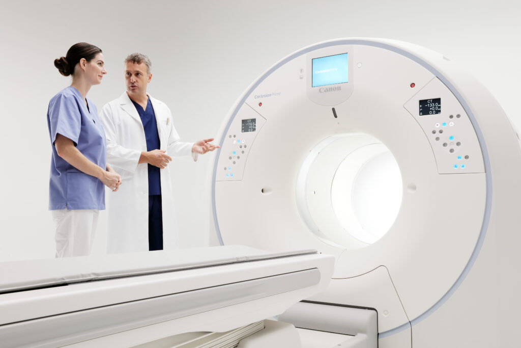
MRH’s newest CT scanner, the Cartesion Prime Digital PET/CT from Canon Medical Systems, features detailed imagery, faster scan times, and even AI-assisted technology.
“We are always striving for the best imaging technology to provide better results for our patients,” says Bodie Blowers, Director of Medical Imaging at Montrose Regional Health.
Learn more about this advanced CT technology here.
MRH Medical Imaging provides services for every organ system of the body. Interventional radiologists can treat a wide variety of conditions that once required surgery.
This is performed by using catheters and other tiny instruments, guided by radiologic imaging. The advantages of interventional radiology include reduced risk, pain and recovery times, and less expensive than surgical alternatives.
Magnetic Resonance Imaging (MRI) is an imaging tool that produces very detailed images without the use of X-rays. By using a large magnet, radio waves and a computer system to process the data, images are created without involving ionizing radiation.
In the typical MRI exam, you will be asked to lie down in the scanner and a device — called an imaging coil — will be placed over the area of your body to be examined. It is important for you to be able to lay still during the process, since movement can result in poor image quality. The process may involve taking a series of images, each series lasting approximately 10-15 minutes. 2-3 series may be needed depending on the situation. Please contact us if you have any questions about your exam.
Our equipment produces high-quality images—an indispensable resource for physicians in the planning of their patients’ medical treatment.
Safety Screening Questionnaire
Because an MRI examination involves the use of a very strong magnet, you will be asked to fill out a safety screen questionnaire when you arrive for your safety. Should you need assistance completing this form, technologists will be available to assist you.
In X-ray and CT examinations, external sources use X-ray to produce the images that inform caregivers. Nuclear medicine images are different: they are obtained by injecting patients with radiotracers or by administering the tracer orally. Through the normal, physiological processes of the body, the substance localizes to a particular site of the body — allowing for a gamma camera to image the location and provide information about what is going on in the body.
In addition to the standard nuclear medicine examinations, MRH also offers PLANAR and SPECT imaging: advanced nuclear imaging technology. Each situation will dictate which method will be used for the care of you or your loved one.
An ultrasound examination is a safe procedure that produces an image using very high-frequency sound waves. Since the ultrasound machine’s development in the 1950s, technology has improved to the point that images are now of outstanding clarity and detail.
What are the benefits of having an ultrasound exam instead of an X-ray or CT scan?
Ultrasound uses sound waves instead of X-rays. While X-ray technology produces highly valuable information, sometimes the radiation involved for patients can be avoided by utilizing ultrasound technology instead.
Various studies have indicated that the sound waves used with Ultrasound are completely safe, even on pregnant women (when CT or X-rays would be inappropriate). In addition, ultrasounds are often performed more quickly and with less expense. Your physician will order the best exam for your situation.
Medical X-rays identify bony and soft tissue structures. The most common medical use of X-rays is the Diagnostic X-Ray, which examines the body using basic x-ray techniques.
There are two types of X-ray images, radiographs (still images) or fluoroscopy (“real time” images). Fluoroscopy uses video cameras to visualize internal motion in real-time. Some X-ray exams require either drinking or injecting a “contrast” material to better outline structures to be soon on the image; however, most don’t require any special preparation.
Montrose Regional Health is proud to offer DEXA scans for our communities! DEXA scans are often used to provide details about bone fractures and also your risk for osteoporosis.
According to the CDC, osteoporosis affects almost 20% (1 in 5) of women over 50 and almost 5% (1 in 20) of men over 50. DEXA scans are the gold standard for diagnosing osteoporosis and fracture risk. Ask your provider if a DEXA scan may help assess your bone health today. Then, talk about steps you can take to slow bone loss or protect your bones for years to come.